Contents
Anatomy


General information
- This section is strictly limited to anatomy, you might be looking for clinical relevant information which is found under the clinical chapters -- muscles section, click here to go to that page
Position
- Deep
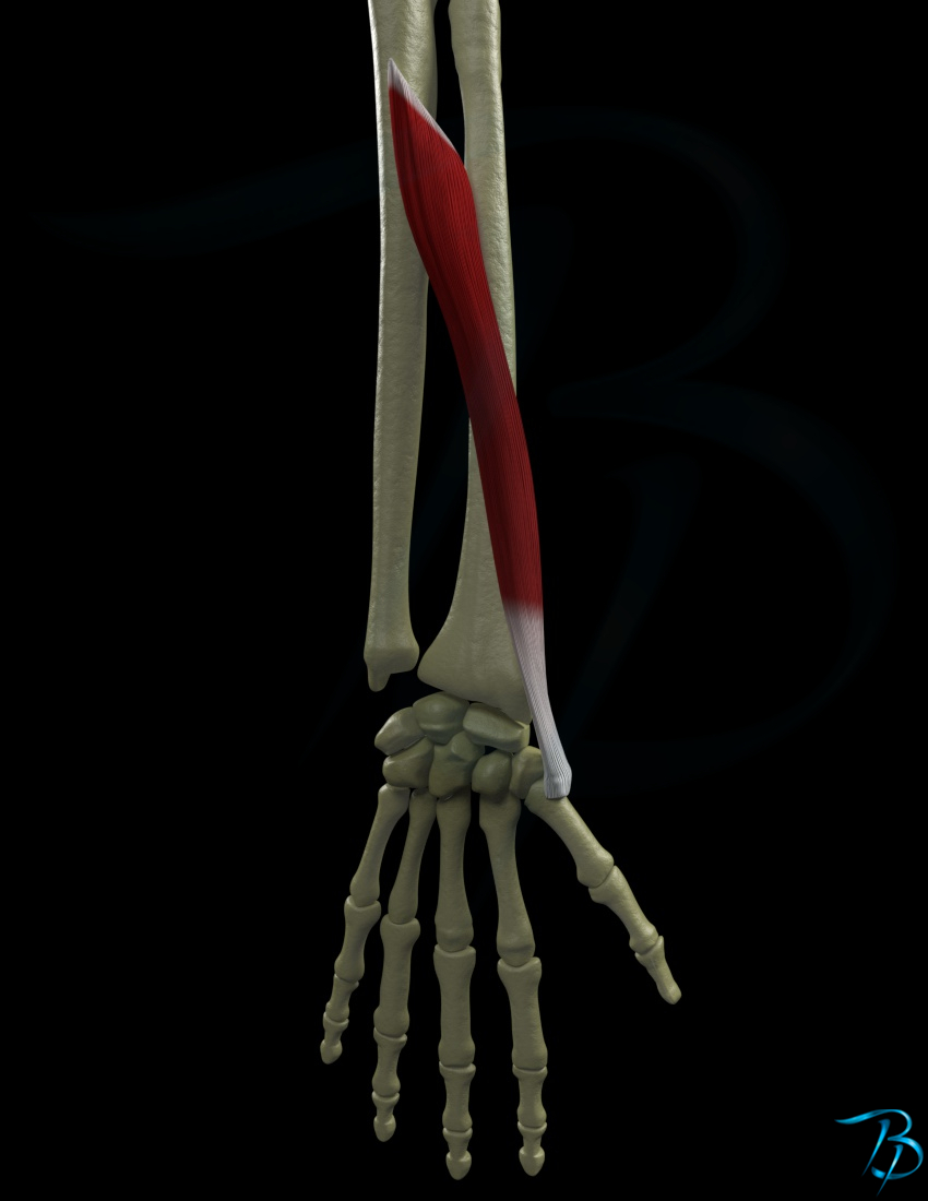
Origin
- Middle 1/3 of radius (posteriorly)
- Middle 1/3 of ulna (posteriorly)
- Middle 1/3 of membrana interossues
Insertion
- Base of 1.metacarpal (radial side)
Main function
- Thumb - CMC
- Abduction
Secondary function
- RU-joint
- Supination
- Wrist
- Radial deviation
- Flexion
- Thumb - CMC
- Extension
- Lateral rotation
Nerve innervation
- Segmental
- C6-C8
- Peripheral
- Nervus radialis
Arterial supply
- Posterior interosseus artery
- Anterior interosseus artery
Palpation
- Pasient position: Sitting
- Ask the patient to hold the radioulnar joint in a neutral position between pronation and supination
- Place your hand on the lateral aspect of the wrist of the patient
- Ask the patient to abduct and extend the thumb while you feel for the tendon of abductor pollicis longus and extensor pollicis brevis
- Palpate along the muscle in a distal direction until you find the insertion of the 1.metacarpal.
- At this position you can seperate the two tendons with a finger nail, the most anterolateral part of these is the abductor pollicis longus
Strength test
- Patient position: Sitting or supine
- Stabilize the patient's wrist
- Ask the patient to hold the thumb in an abducted and extended position
- Give resistance towards the lateral and distal side of the 1.metacarpal in the direction of adduction and flexion, so that the patient is giving force towards abduction and extension