The anterior surface is fascially continous to the iliacus
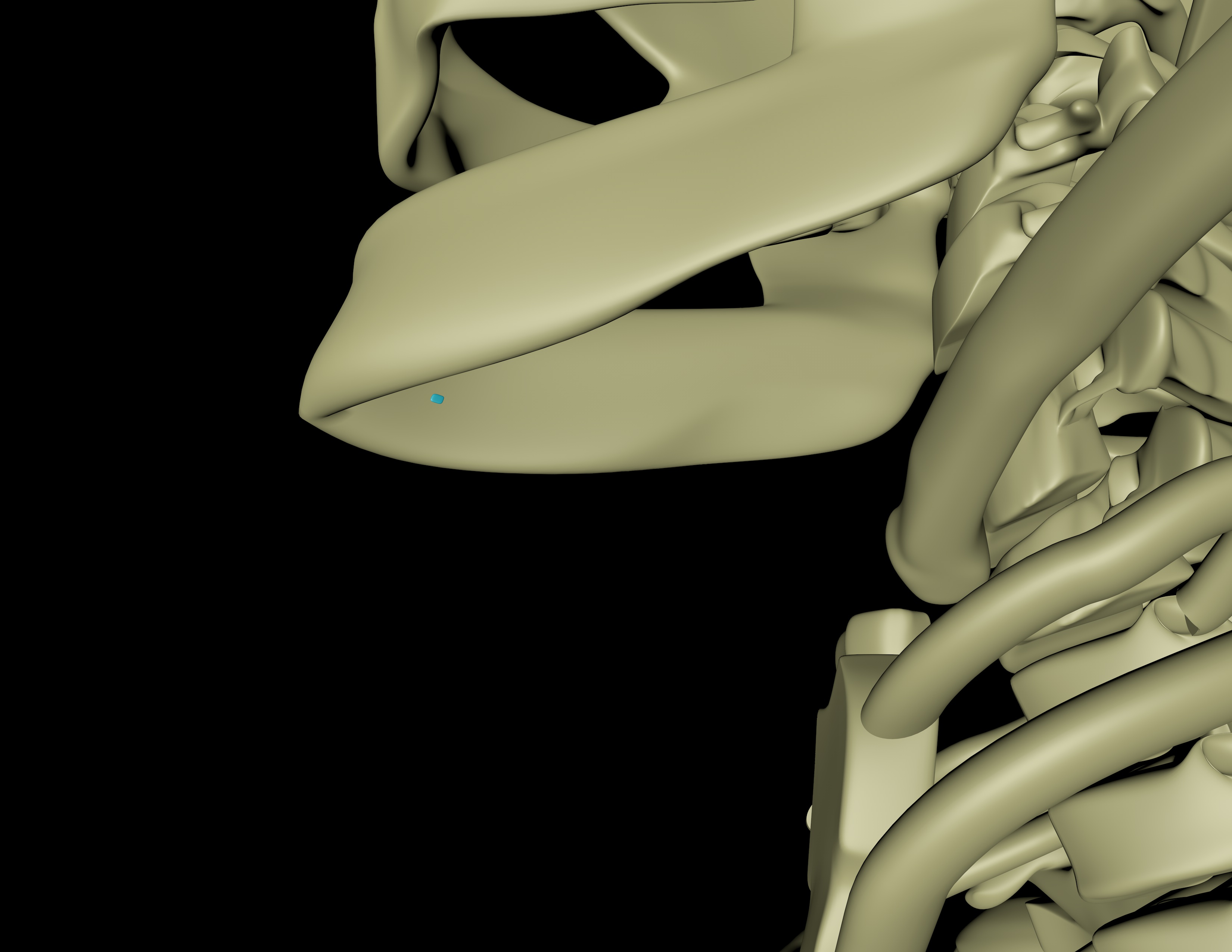
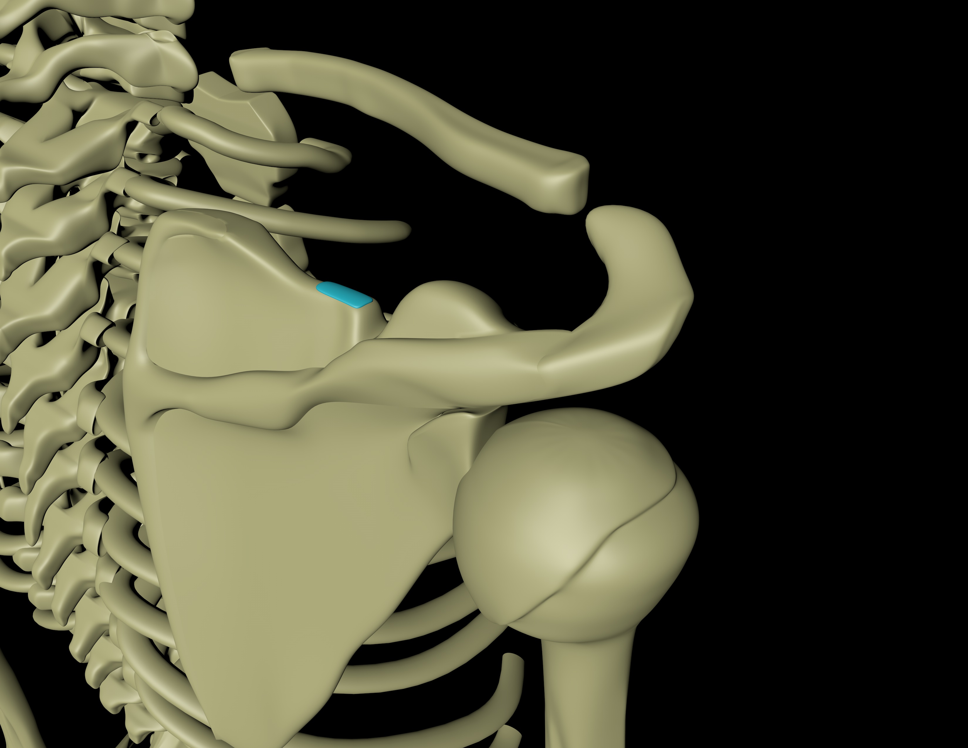
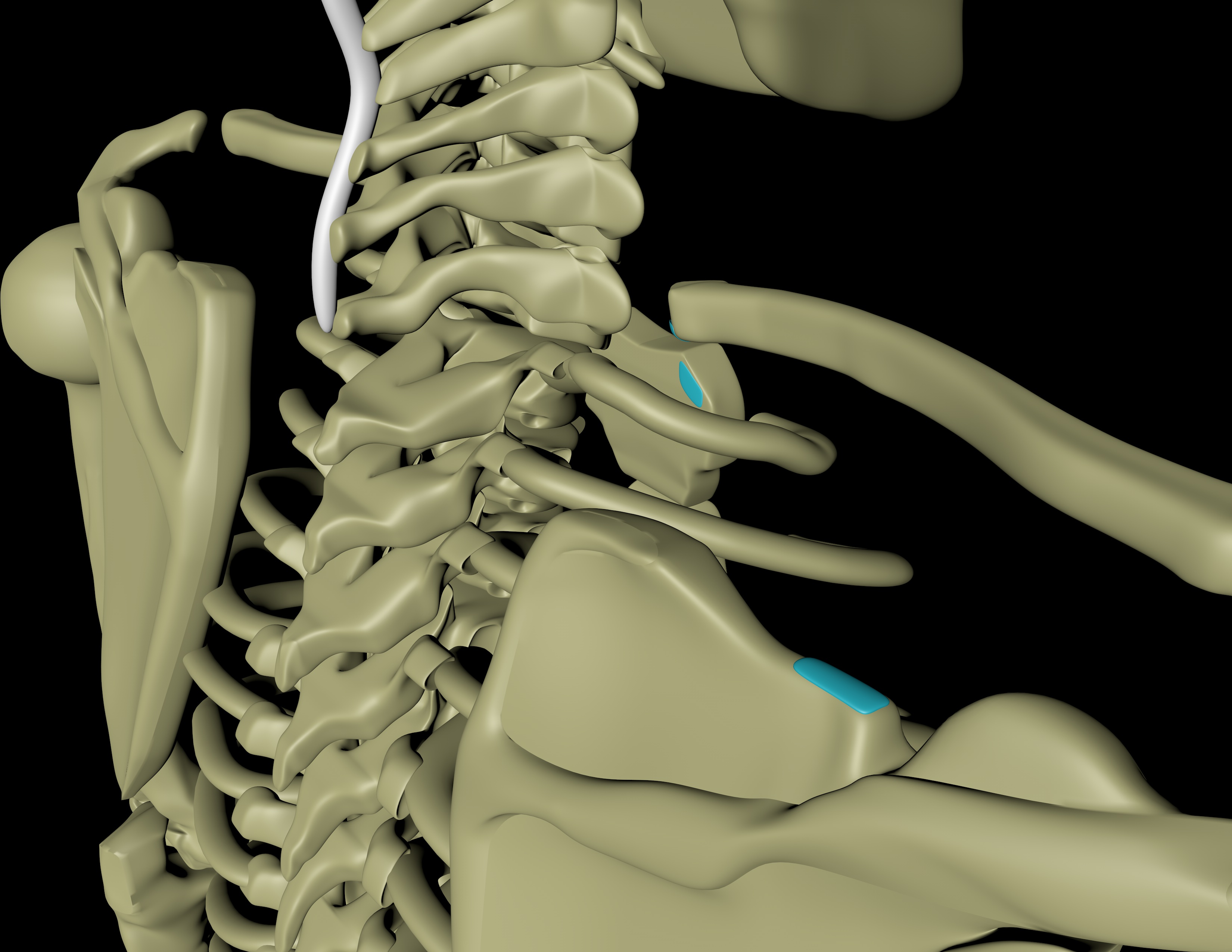
Attaches To The Following Bones
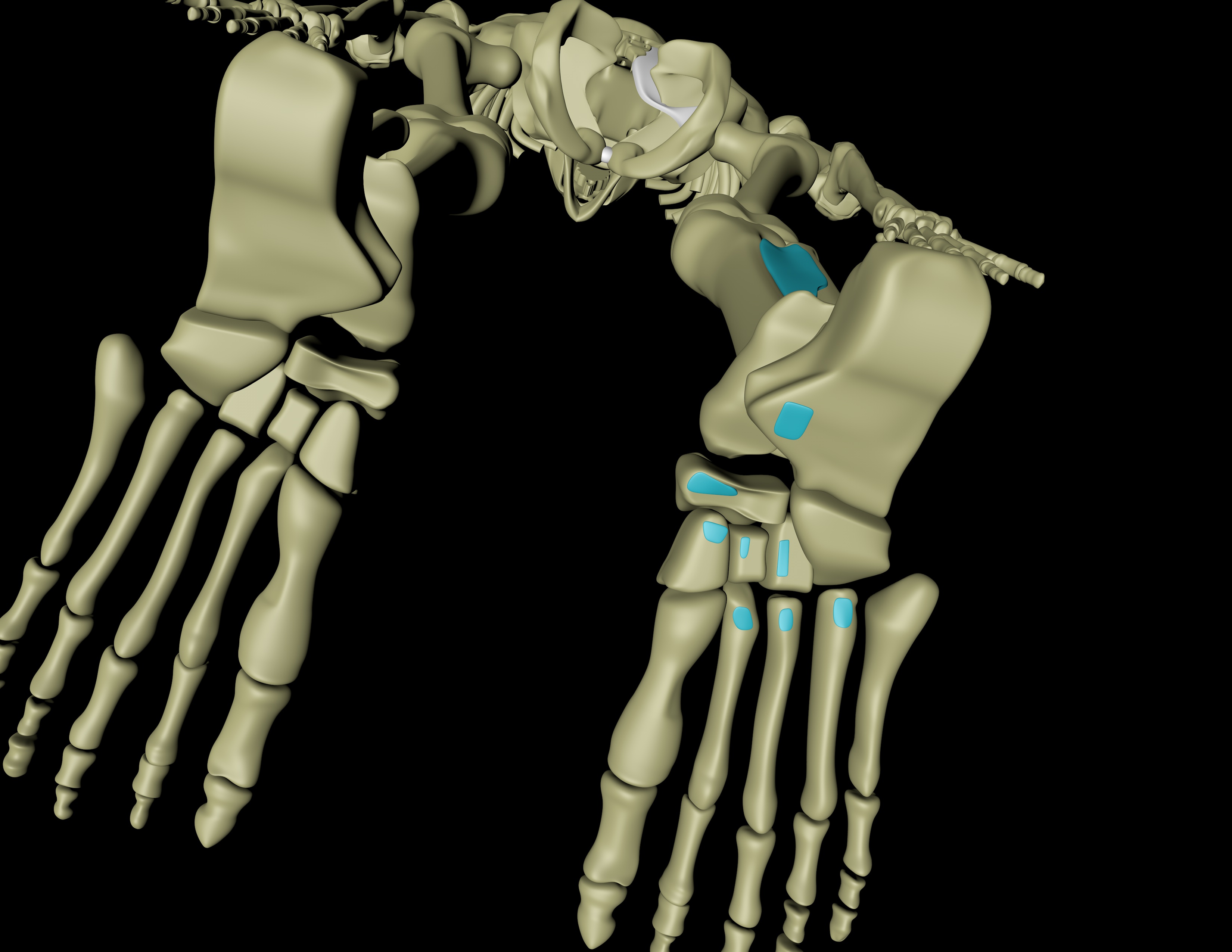
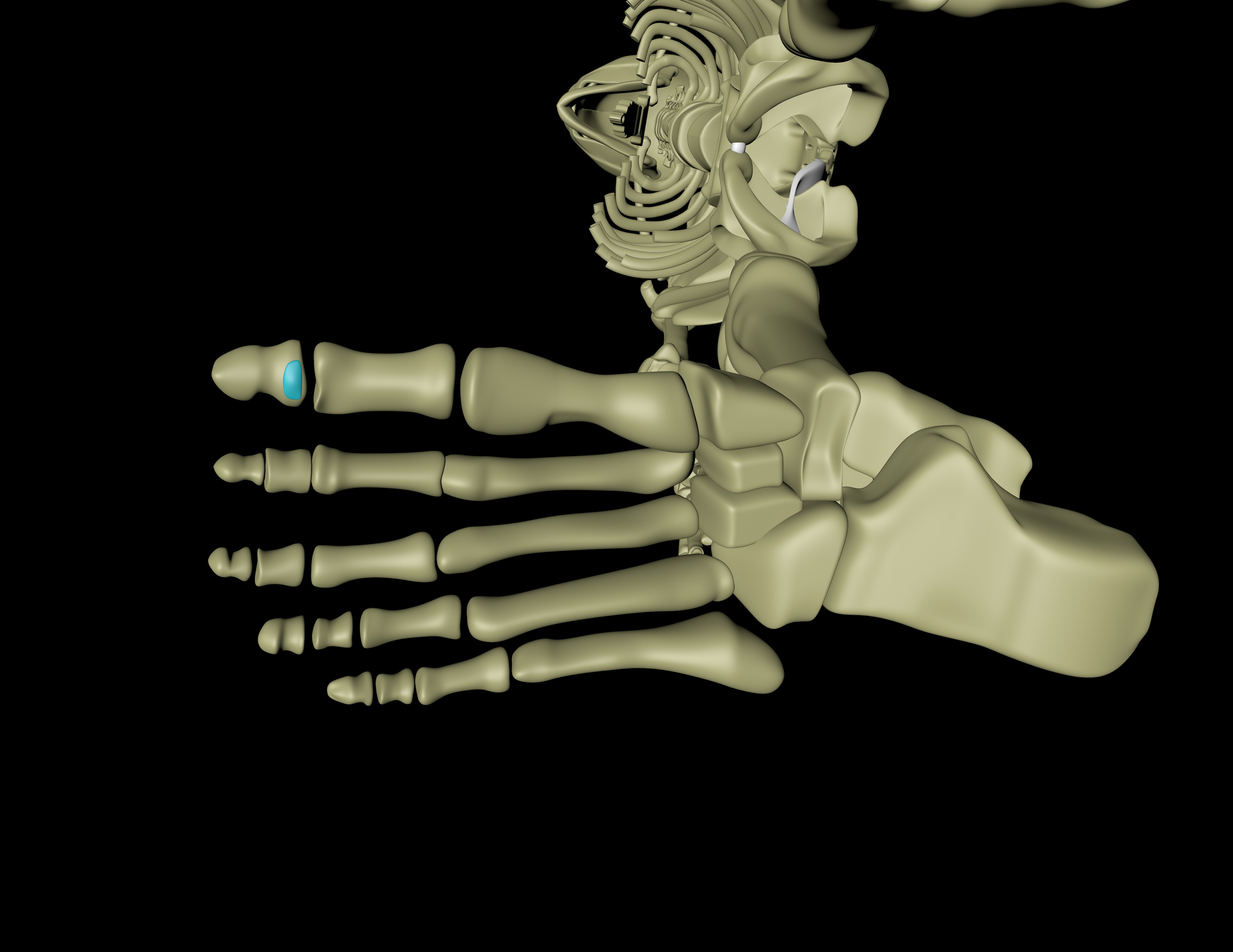
Tarsal plantar bones, plantar surface of toes
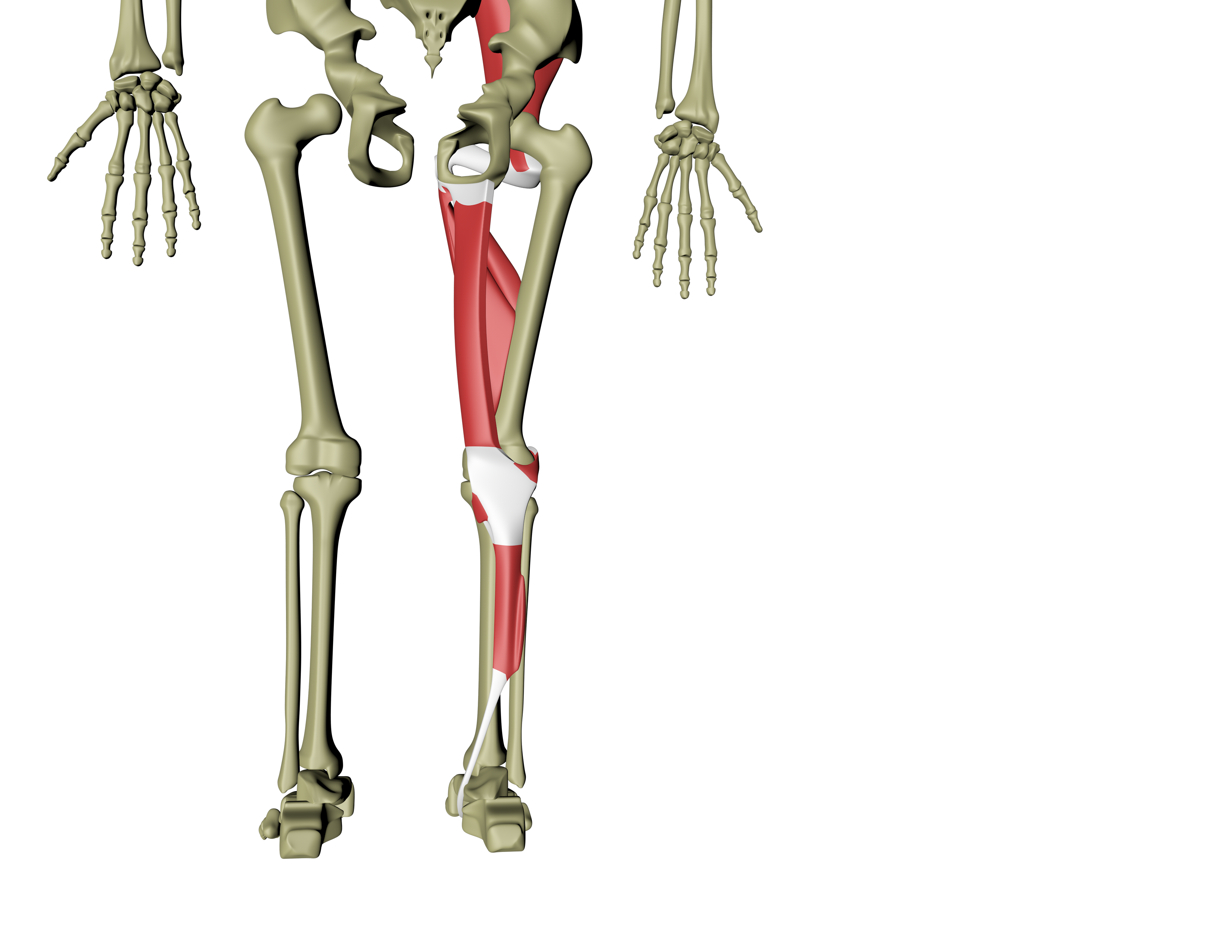
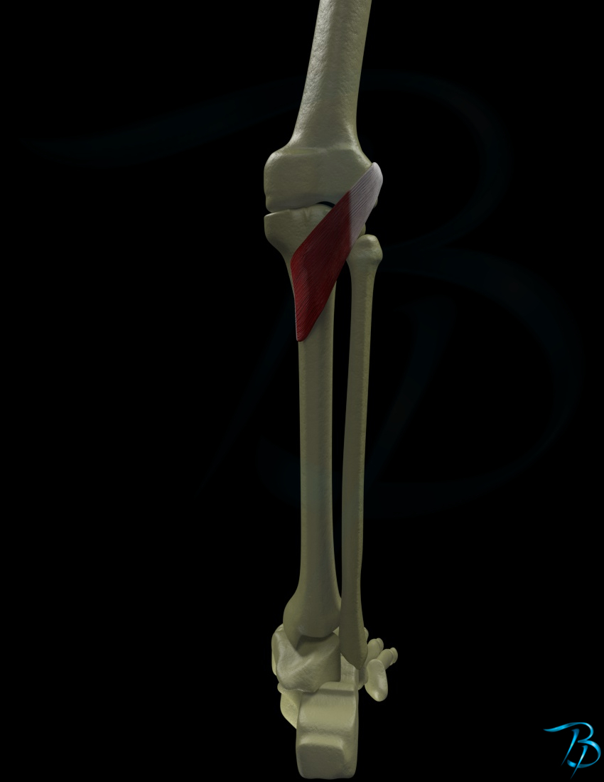
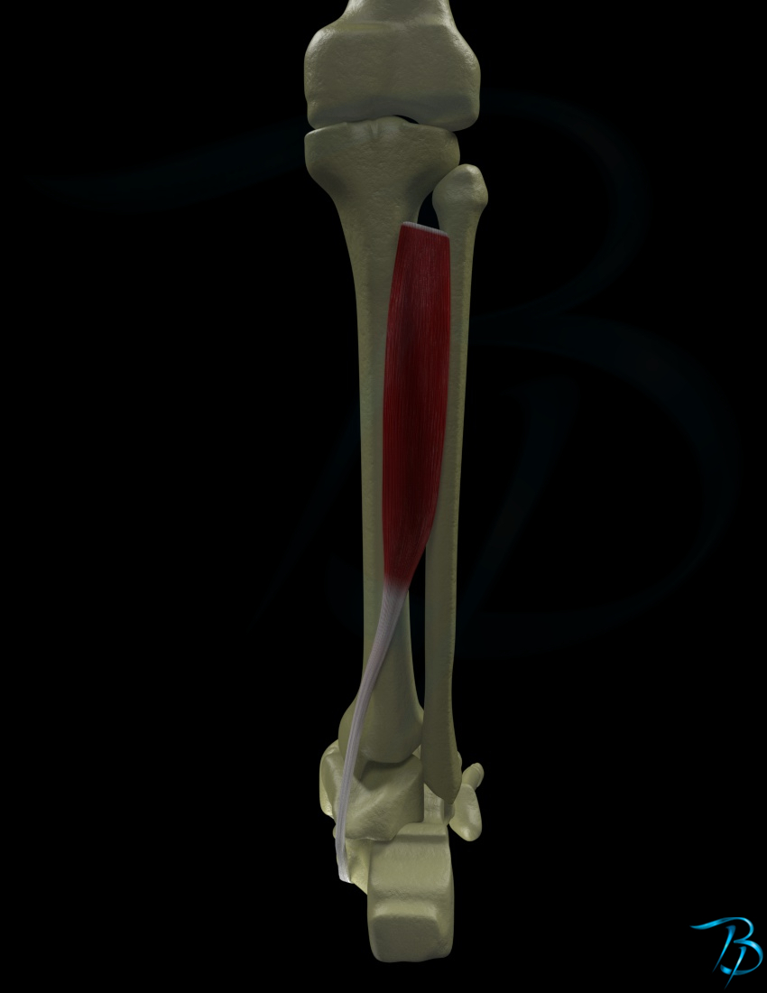
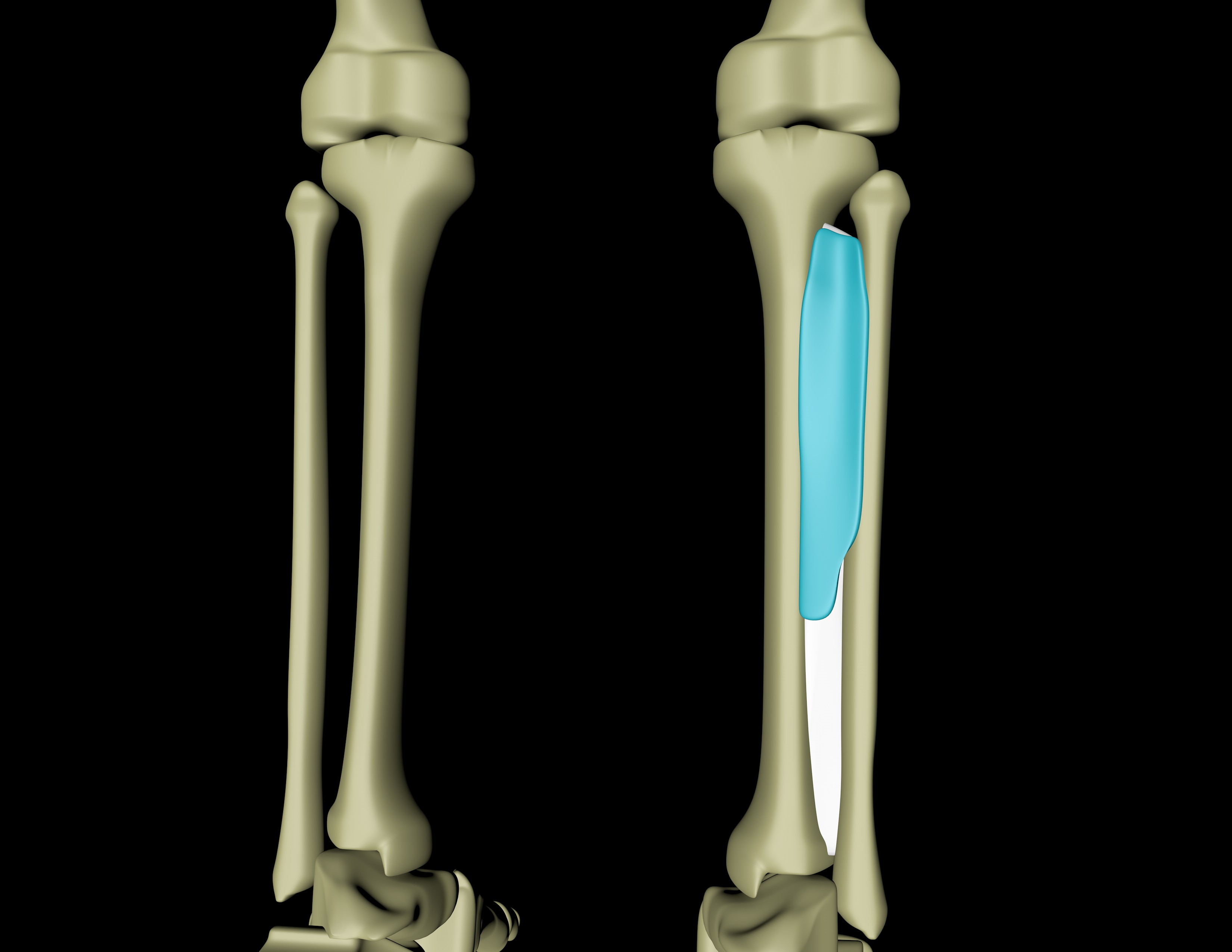
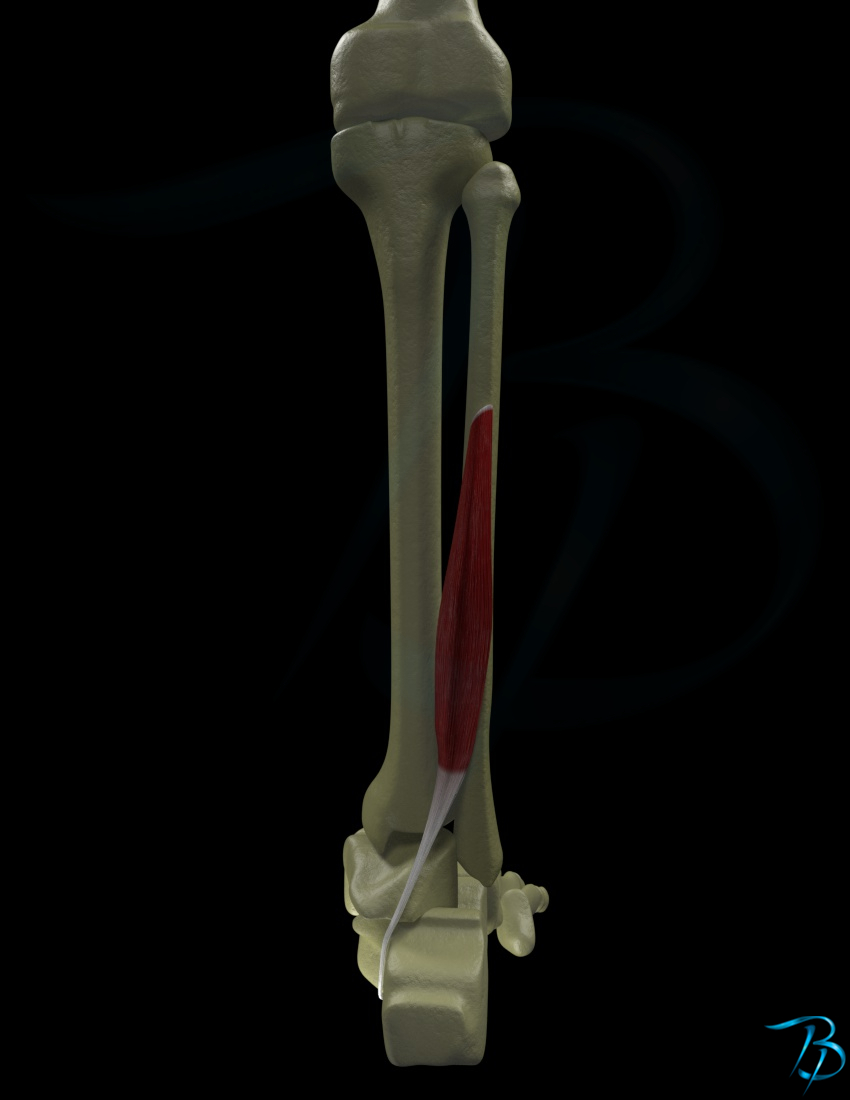
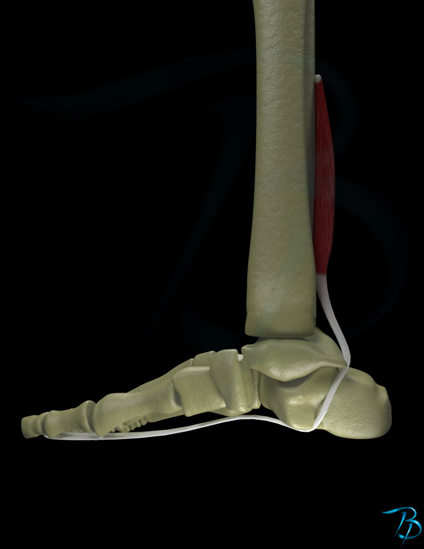
Superior/posterior tibia/fibula
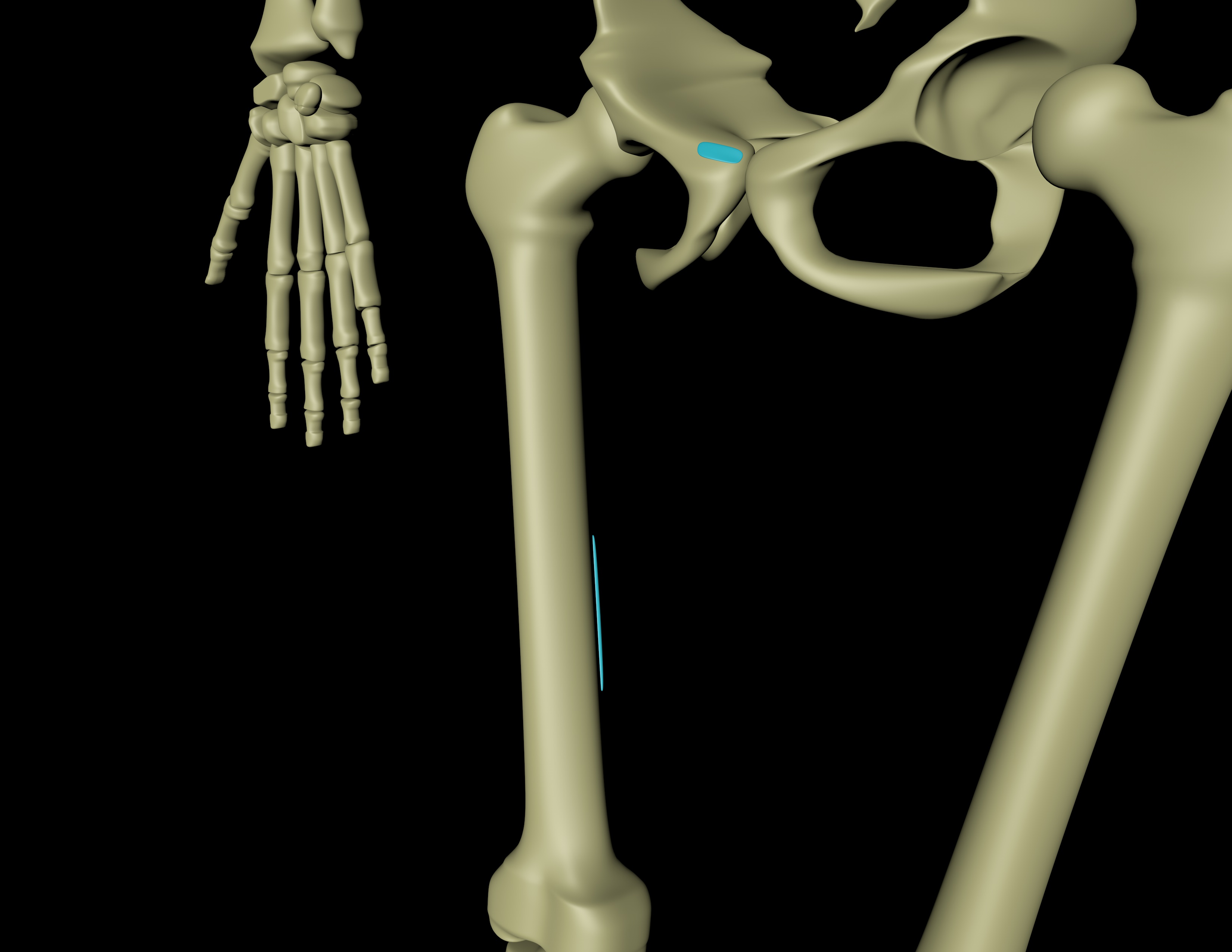
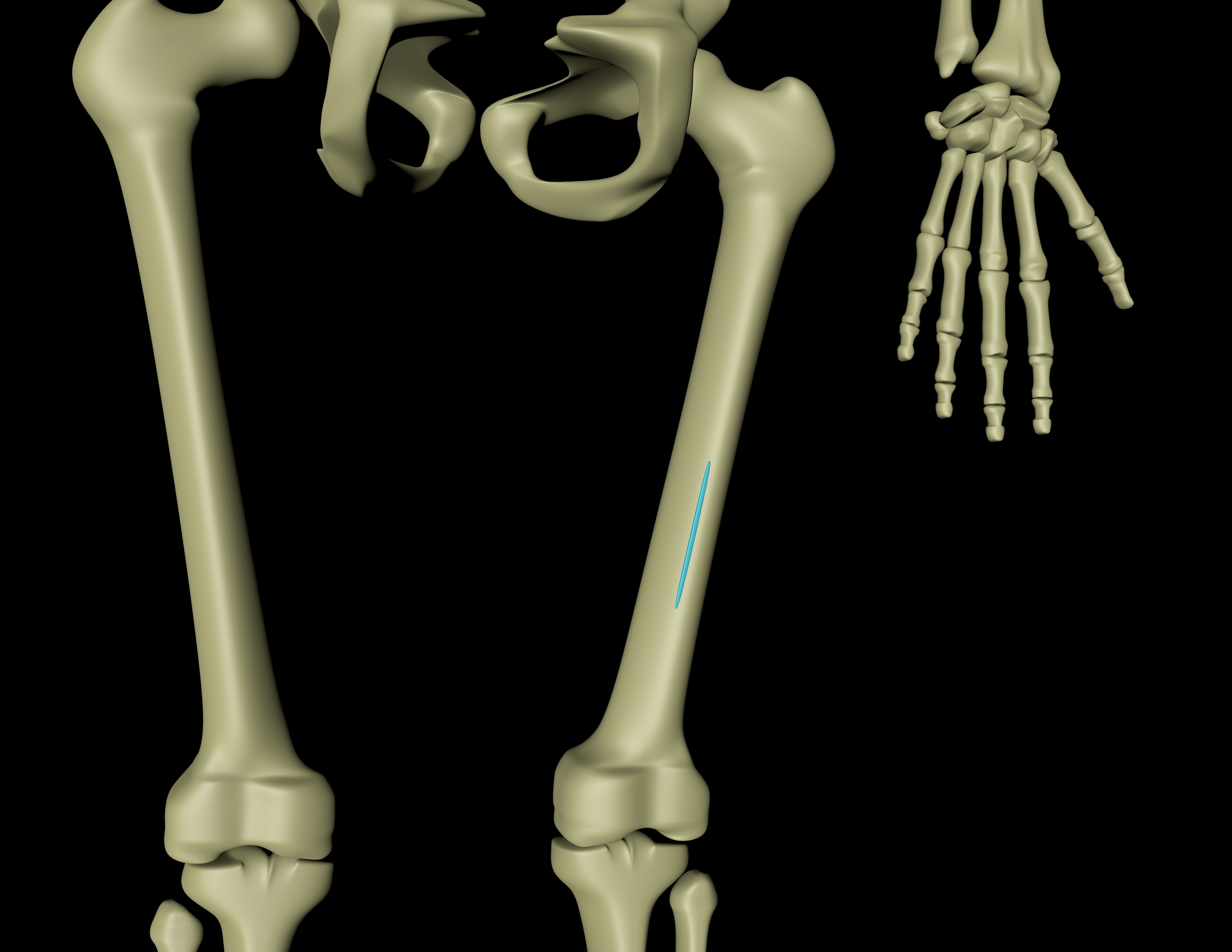
Medial femoral epicondyle
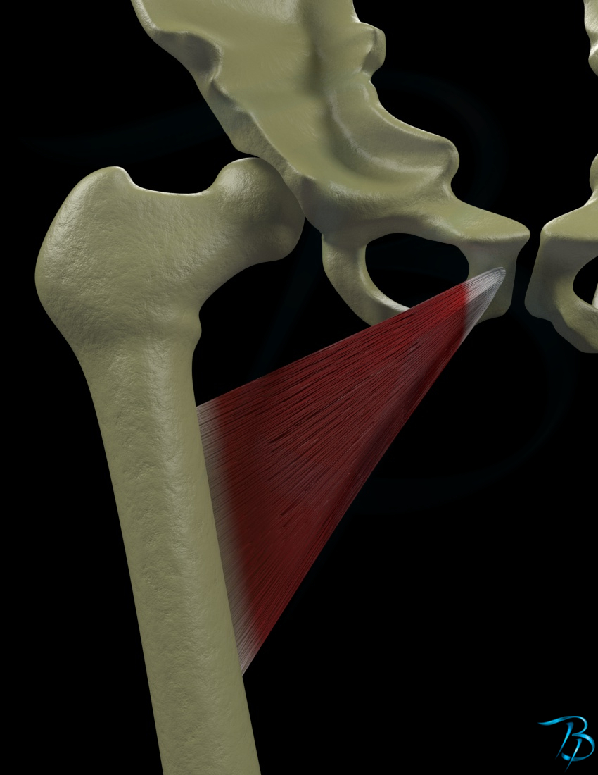
Linea aspera femoris
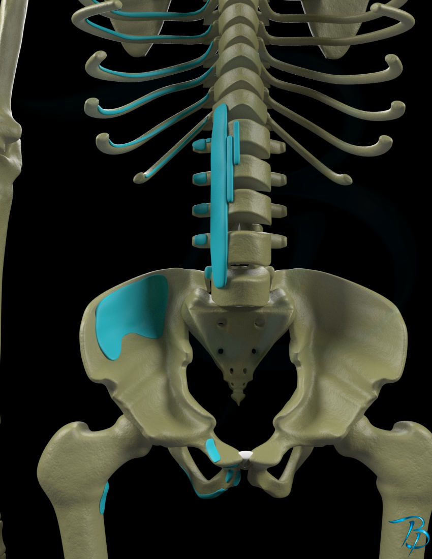
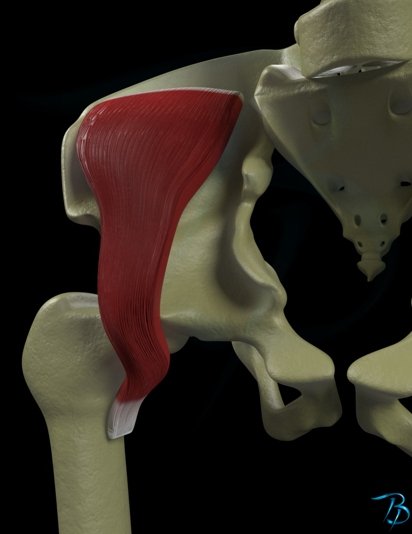
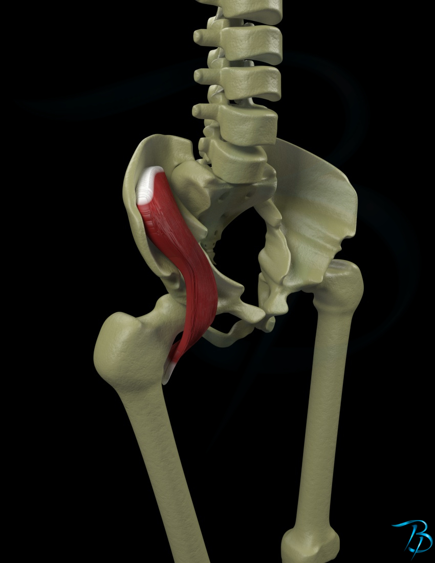
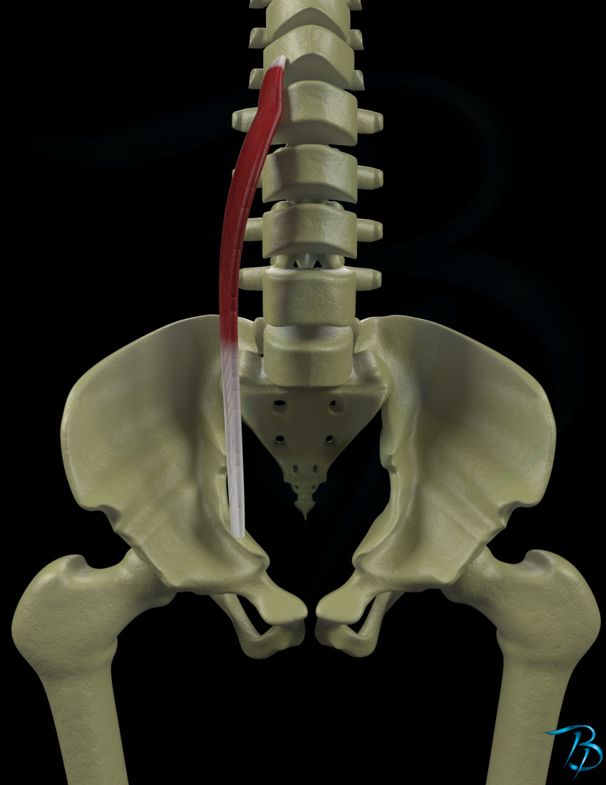
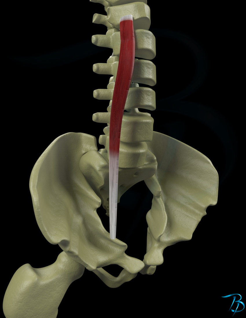
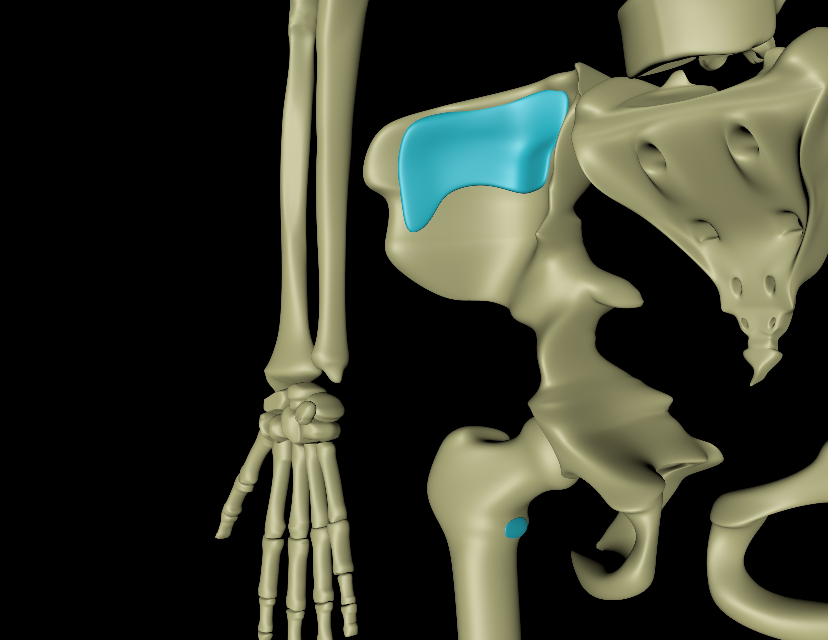
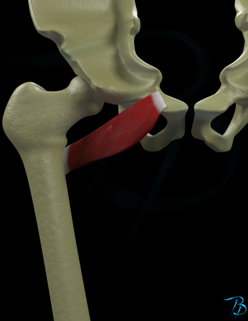
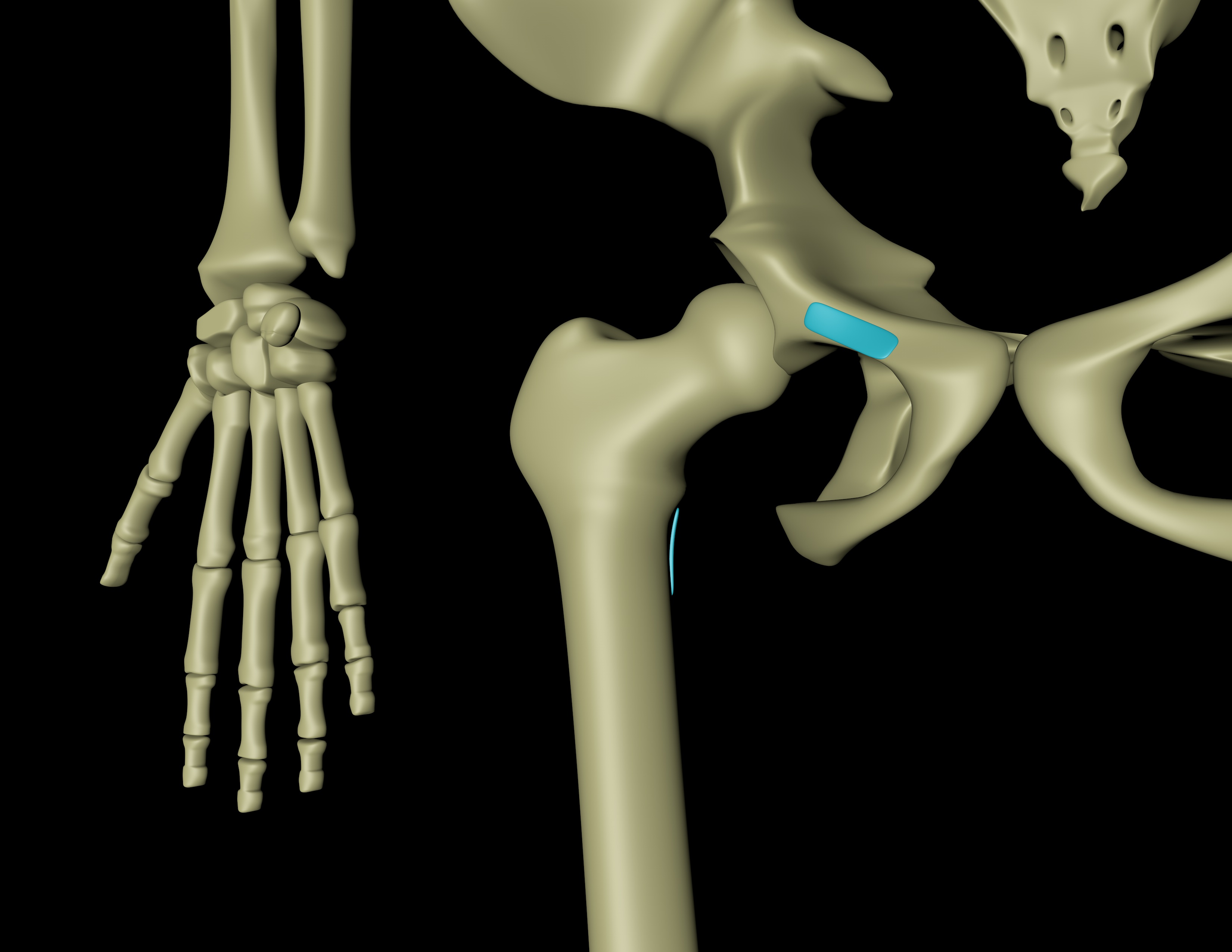
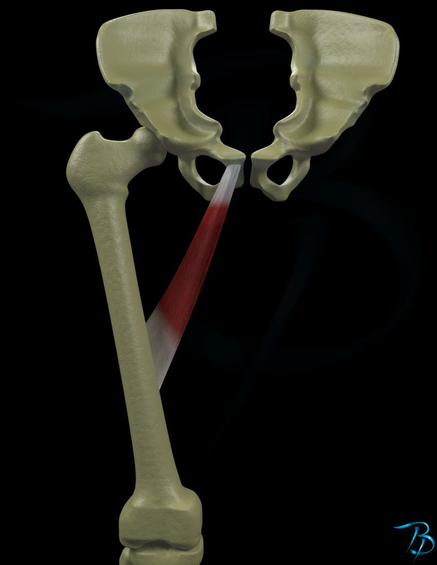
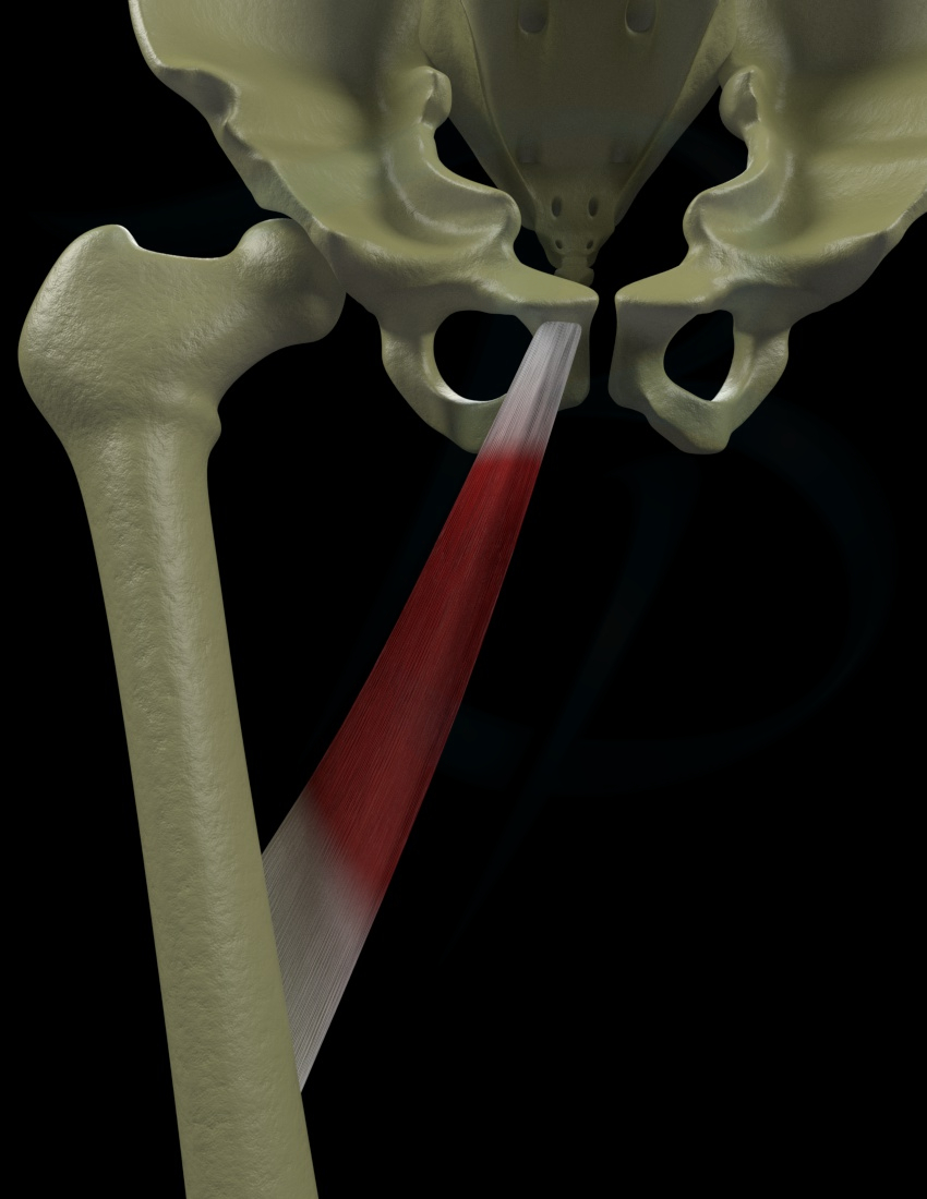
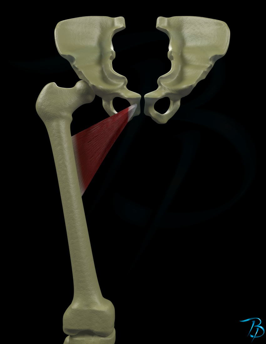
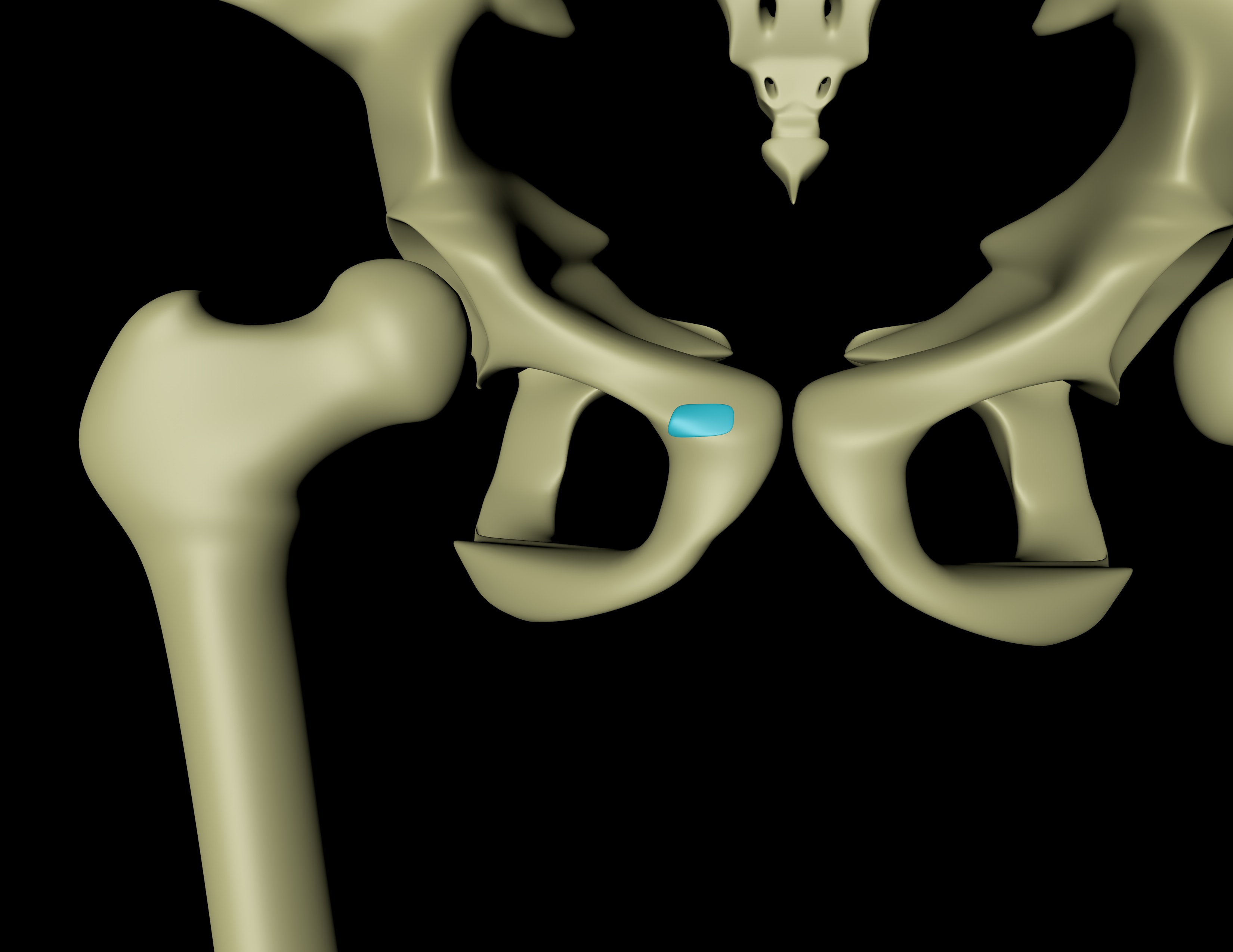
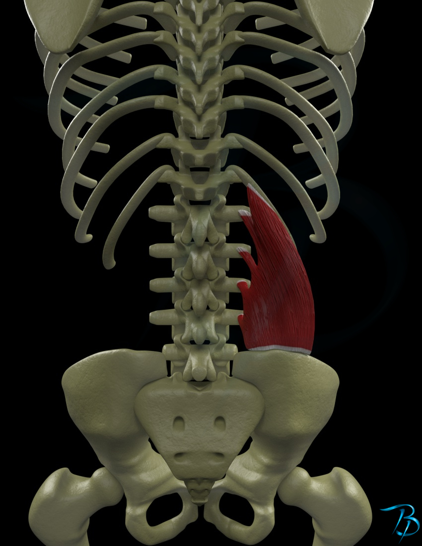
Trochanter minor femoris
Ischial ramus
Coccyx
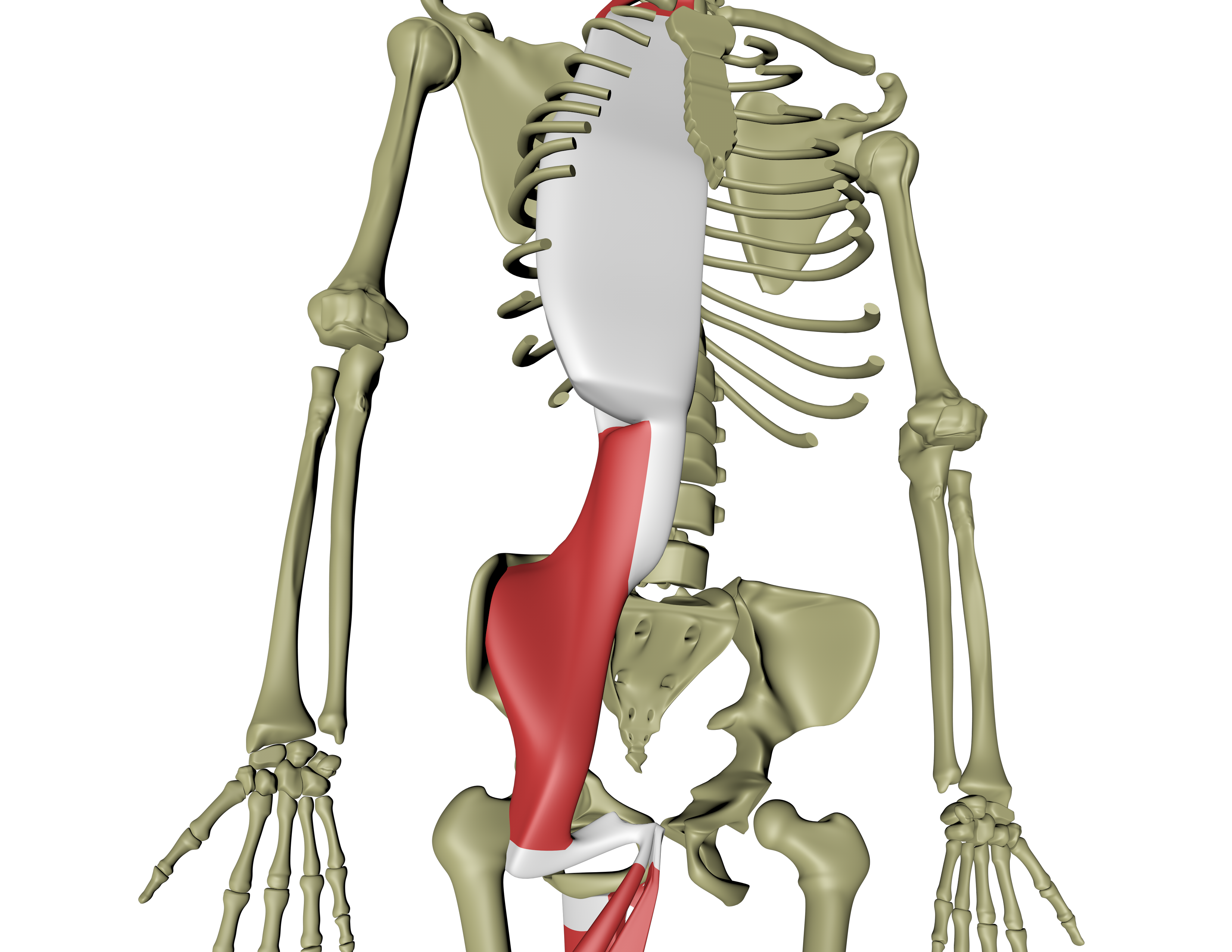
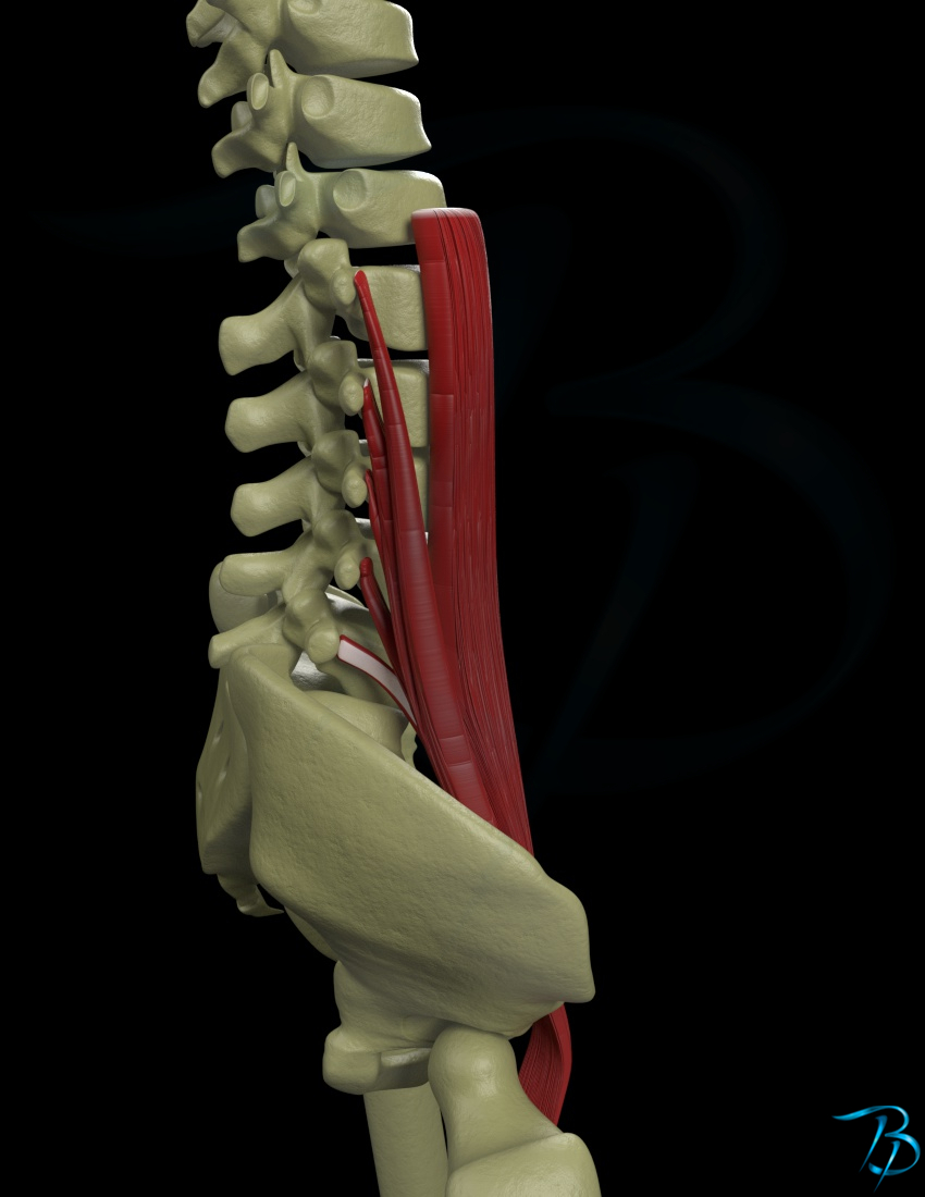
Lumbar vertebral bodies and processus transversi
Posterior surface of subcostal, cartilages and processus xiphoideus
Posterior manubrium
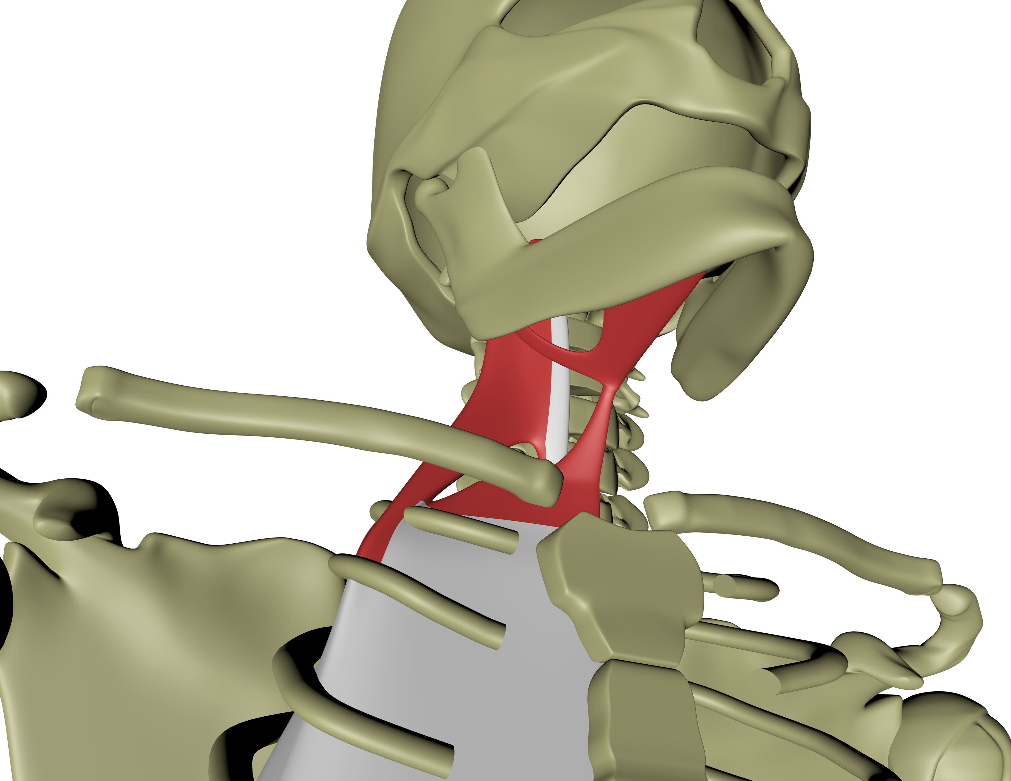
Hyoid bone
Mandible
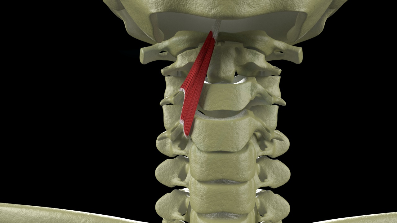
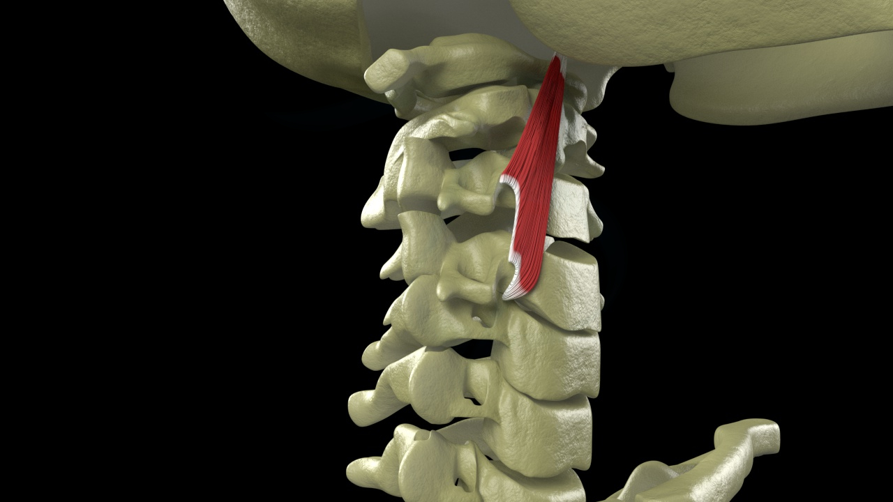
Processus transversi of cervicals
Basilar portion of occiput
Function of line
Lifting the inner arch of the body, and therefore important in the body's support
Giving support to the lumbar spine from the front
Stabilizing the chest while allowing the expansion of the ribs for relaxation of breathing
Stabilizing and balancing the neck and head
Stabilizing each segment of the legs
Overload of the line will result in less bodily stabilization and higher involvement of the other lines to support the body. Thus increasing the load of other muscles and structures and simultaneously increasing the likelihood of causing injuries to these compensated lines
Compensation Patterns Associated With The Line
Chronic plantarflexion
High and fallen arch patterns of the feet
Pronation and supination
Genu valgus and varus
Anterior pelvic tilt
Pelvic floor insufficiency
Lumbar malalignment
Breathing restriction
Flexed or hyperextended cervicals
Temperomandibular joint syndrome (TMJ)
Swallowing and language difficulties
General core collapse
Dysregulation of line
Produces overall shortening in the body, encourage collapse in the pelvic and spinal core
Eventhough the line is dysregulated, in many cases there may be several years before a obvious injury occurs due to the body's ability to compensate. So a loss of function within the line does not necessarily involve an immediate injury or collapse