Contents
Anatomy


General information
- This section is strictly limited to anatomy, you might be looking for clinical relevant information which is found under the clinical chapters -- muscles section, click here to go to that page
Position
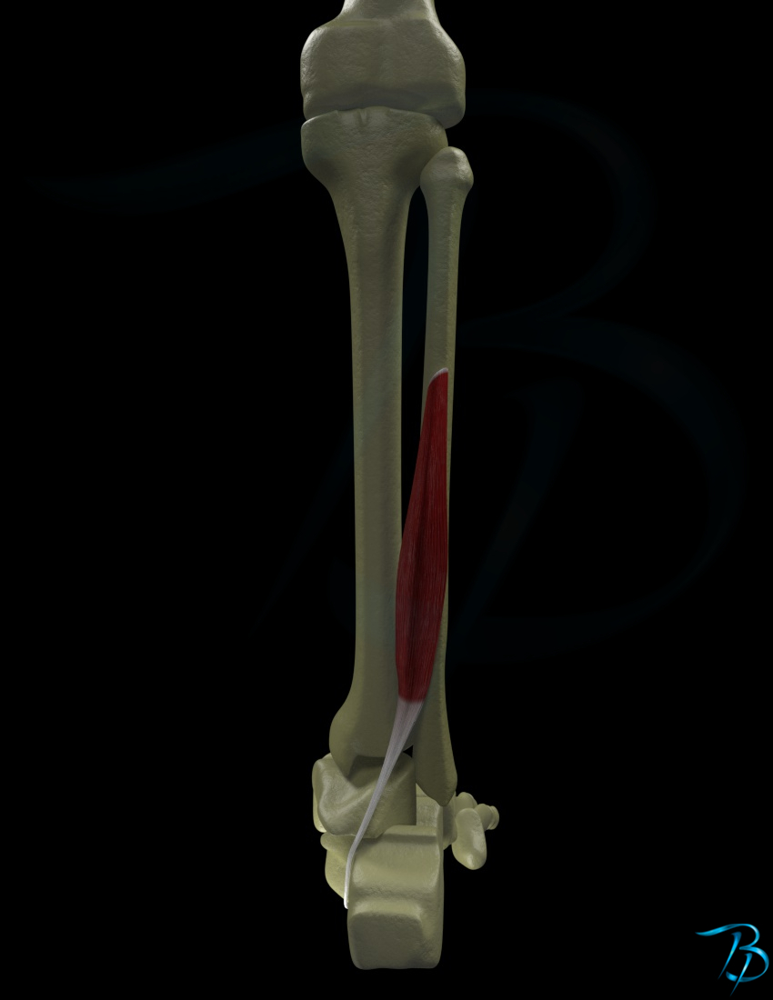
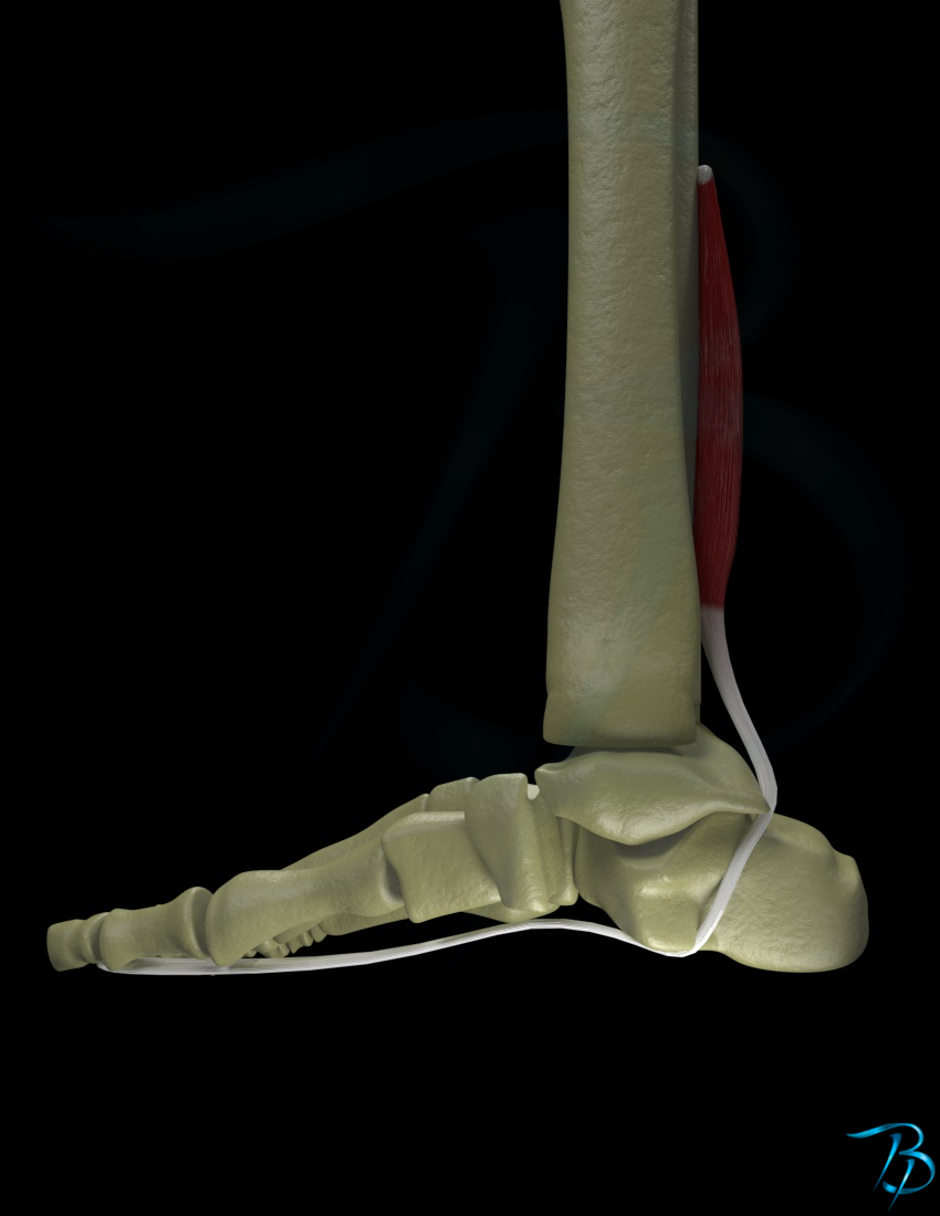
- Deep
Origin
- Posterior surface of distal 2/3 of fibula
- Membrana interosseus
- Intermuscular septum and fascia
Insertion
- Base and plantar side of distal phalanx at metatarsal 1
Main function
- IP-joint
- Flexion
Secondary function
- Ankle
- Plantarflexion
- Tarsal-joint
- Inversion
- MCP-joint
- Flexion
Nerve innervation
- Segmental
- L5-S2
- Peripheral
- Tibial nerve
Arterial supply
- Posterior part of tibial artery
Palpation of distal tendon
- Patient position
- Supine
- Place your hand just posteriorly to the medial malleol
- There are 3 tendons running down posteriorly to the medial malleol:
- Tibialis posterior
- Flexor digitorum longus
- Flexor hallucis longus
- Ask the patient to flex the big toe while you feel for the contraction in the tendon of flexor hallucis longus
Strength test
- Patient position
- Supine
- Staiblize the patient's foot by holding around the metatarsal bones while the foot is held in a neutral position
- Give resistance towards the plantar side of the distal phalanx of the big toe in an extension direction, so that the patient is giving force towards flexion