Contents
Anatomy


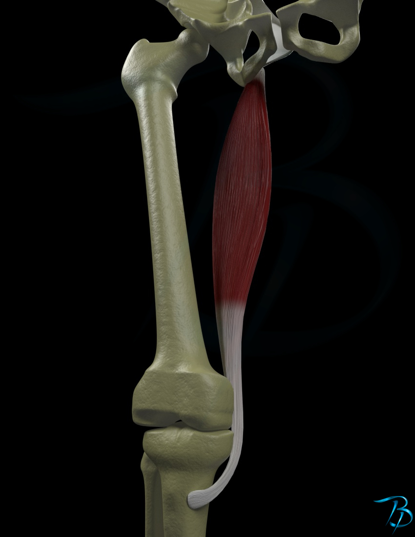

General information
- This section is strictly limited to anatomy, you might be looking for clinical relevant information which is found under the clinical chapters -- muscles section, click here to go to that page
Position
- Superficial
Origin
- Tuberositas ischiadicum
- Via the common tendon with biceps femoris
Insertion
- Pes anserinus
- Body of tibia
- Proximal and medial surface
- Deep fascia of the calf
Main function
- Knee
- Flexion
- Hip
- Extension
- Pelvis
- Posterior tilt
Secondary function
- Knee
- Medial rotation
- Hip
- Medial rotation
Nerve innervation
- Segmental
- L5-S2
- Peripheral
- Sciatic nerve
Arterial supply
- Ingerior gluteal artery
- Deep part of the femoral artery
- Popliteal artery
Palpation
- Patient position
- Prone
- Place a chair or similar object on the treatment bench that the patient can use to rest their foot on, so that the knee is flexed
- Place your hand posteromedially and distally on the patient's thigh
- Use your opposite hand to give resistance towards the patient's heel, so that the patient is giving force towards flexion
- Feel for the contraction of the muscle
Strength test
- Patient position
- Prone
- Fixate the patient's thigh down towards the bench with your one hand
- Bend the patient's knee to about 60°
- Position the hip so that it is in slight medial rotation
- Position the knee so that too is in slight medial rotation
- Give resistance to the patient posteriorly and distally at the patient's calf, just proximal to the ankle
- The pressure is in extension direction of the knee so that the patient is giving force towards flexion of the knee
Weakness of
- In weakness, the patient may have problems holding the foot in a rotated position which is the starting point of the test
- Weakness of the medial hamstrings will diminish the medial stability, so that during gait there may be a valgus positoin of the knee, also called 'knock-knee'
- If all of the hamstrings group is weak, this will lead to the knee being hyperextended
- In bilateral weakness of both hamstrings, the pelvis will compensate by tilting anteriorly, so that the patient has a lordosis of the lower back