Contents
Anatomy

General information
- This section is strictly limited to anatomy, you might be looking for clinical relevant information which is found under the clinical chapters -- muscles section, click here to go to that page
- Tightens the fascia latae
Position
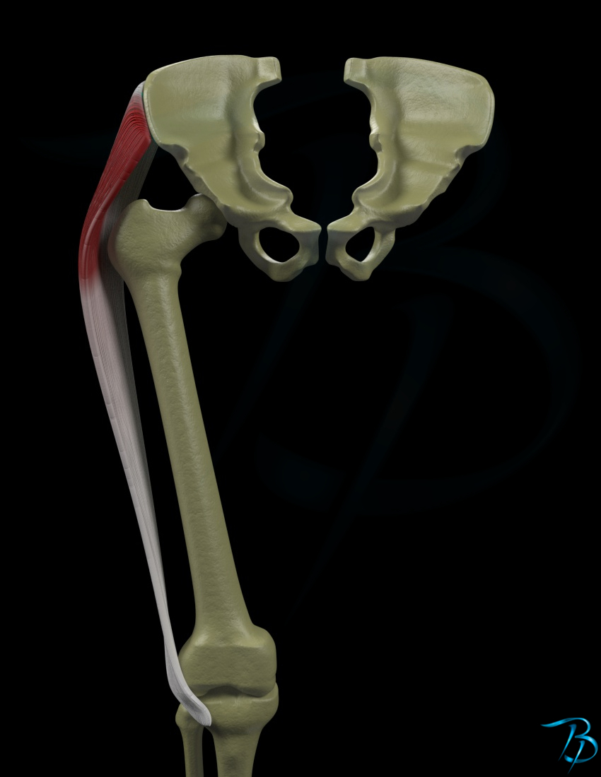
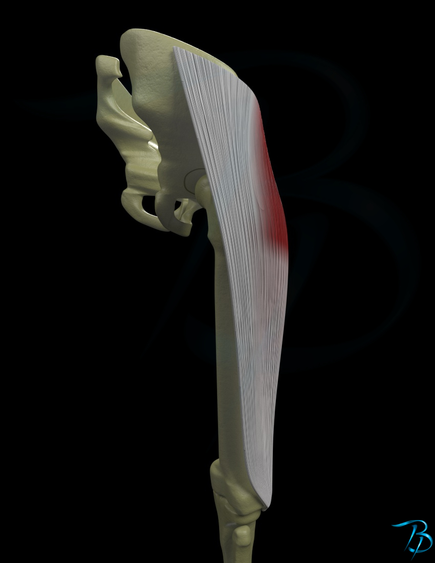
- Superficial
Origin
- SIAS (Spina iliaca anterior superior)
- Anterior part of the ilium crest
- Deep surface of the lata fascia
Insertion
- Iliotibial band
- From proximal 1/3 to the middle 1/3 of it's length
Main function
- Hip
- Flexion
- Abduction
- Medial rotation
- Pelvis
- Anterior tilt
Secondary function
- Hip
- Depression
- Pelvis
- Ipsilateral rotation
- Knee
- Extension
Nerve innervation
- Segmental
- L4-S1
- Peripheral
- Superior gluteal nerve
Arterial supply
- Superior gluteal artery
- Deep femoral artery
Palpation
- Patient position
- Supine
- Palpate the SIAS and place your hand just distally and laterally to this point
- Ask the patient to hold the leg in a flexed and medially rotated position of the hip
- Give resistance to the patient in a direction of extension (for the hip) so that the patient is using the flexors while you feel for the contraction of the muscle
- You can palpate the tensor fascia latae distally towards the insertion at the iliotibial band
Strength test
- Patient position
- Supine
- Fixate the patient's pelvis on the opposite side during the testing (if the patient has problems holding the pelvis rested towards the bench)
- Place a pressure towards the patient in a direction of extension and adduction so that the patient is giving force towards flexion and abduction
- The patient's knee is hold in full extension throughout the test
- Weakness can be seen if the patient has problems holding the leg in a medial rotation throughout the test
- Test bilaterally to notice any differences, bilateral weakness is not uncommon