Contents
Anatomy


General information
- This section is strictly limited to anatomy, you might be looking for clinical relevant information which is found under the clinical chapters -- muscles section, click here to go to that page
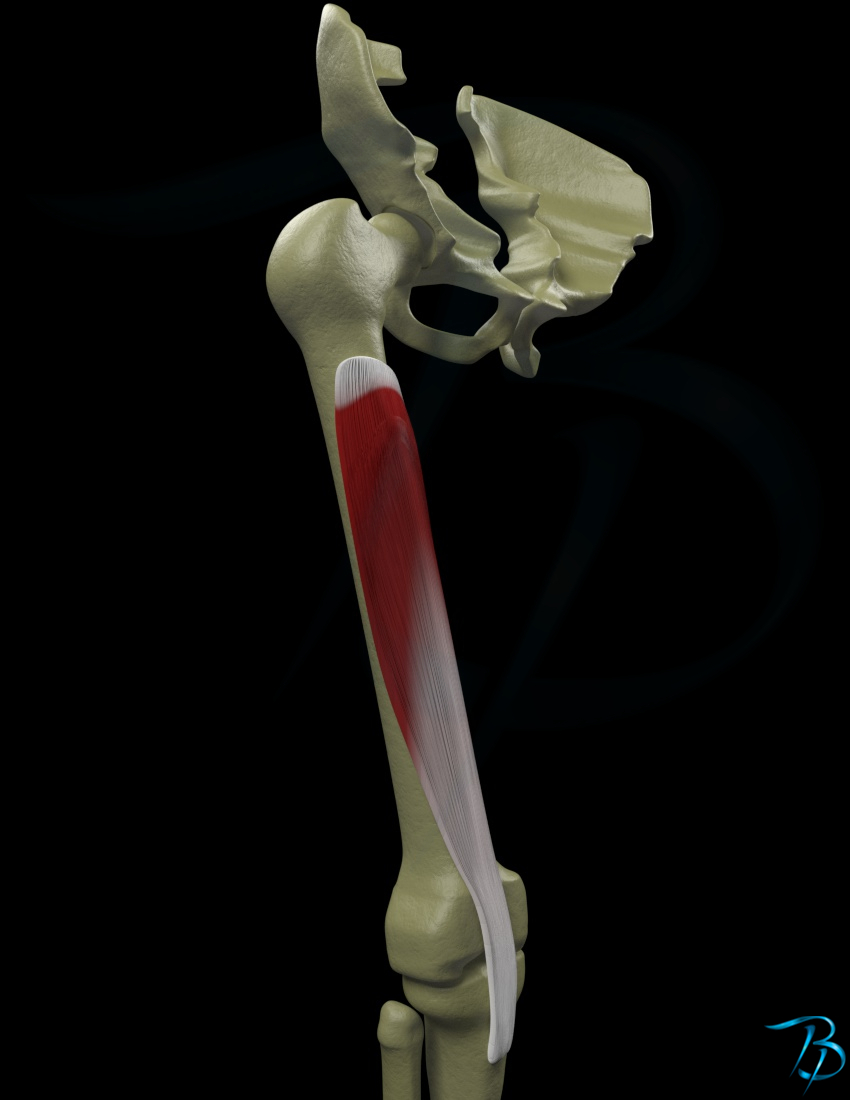
- The deepest of the 4 quadriceps muscles
Position
- Deep
Origin
- Anterior and lateral surface of the proximal 2/3 of the femur body
- Distal 1/2 of linea aspera
- Lateral septum intermusculare
Insertion
- Tuberoistas tibia
- Via paterlla and patellar ligament
- Proximal limitation of patella
Main function
- Knee
- Extension
Nerve innervation
- Segmental
- L2-L4
- Peripheral
- Femoral nerve
Arterial supply
- Femoral artery
Strength test
- Patient position
- Seated with knees over the edge of the treatment table
- Stabilize the patient's knee by holding your one hand just proximal to the patella ventrally on femur
- Place your other hand just distal and ventral on the tibia just proximal to the ankle joint
- Ask the patient to press the foot out towards extension for the knee while your give resistance and press towards flexion
- Regarding tensor fascia latae
- If the patient is twisting the knee into medial rotation, this may indicate that the patient is trying to compensate by using tensor fascia latae to a larger degree. TFL will also be more active by having the patient lean the upper body backwards during the testing
- Regarding hamstrings
- If the patient is leaning the upper body backwards, then this may also indicate too much pressure on an over-activated hamstring and that the patient is trying to relieve it by having less stretch on the muscle
- Regarding rectus femoris
- A third option as to why the patient compensates by leaning backwards,is that if rectus femoris is stronger than the other quadriceps muscles. In these cases the patient prefers having the hip as extended as possible to have maximum efficiency of the muscle due to leverage